|
Overview
 There are two types of flatfeet. Flexible flatfoot means that the foot has some arch, even if it only appears when the person flexes the feet or stands on the toes. This is a normal condition that is generally painless and does not require treatment. Stiff, inflexible, or painful flatfoot is an abnormal condition and may indicate a bone abnormality in the foot, a disease, or an injury. Flatfeet are a normal condition in infants and toddlers. This is partly the result of fatty deposits along the bottom of the foot that go away as the child grows. It is also because the ligaments in the foot have not fully developed. Flat-footedness in children is generally painless and does not interfere with walking or activity. In fact, as children learn to walk, the soft tissues in the foot tighten and form the arch. Most children develop arches by late childhood. When flatfeet continue into adulthood, most cases are considered normal. Incidence of flatfeet in the general population is unknown. Causes Flat feet can be caused by injury, aging, and weight gain. They can cause pain in the feet and may lead to pain in other parts of the body such as the ankles, knees, or hips. For this reason, it behooves us to treat fallen arches. The question becomes how to do so. Symptoms Fallen arches may induce pain in the heel, the inside of the arch, the ankle, and may even extend up the body into the leg (shin splints), knee, lower back and hip. You may also experience inflammation (swelling, redness, heat and pain) along the inside of the ankle (along the posterior tibial tendon). Additionally, you may notice some changes in the way your foot looks. Your ankle may begin to turn inward (pronate), causing the bottom of your heel to tilt outward. Other secondary symptoms may also show up as the condition progresses, such as hammertoes or bunions. You may also want to check your footprint after you step out of the shower. (It helps if you pretend you?re in a mystery novel, and you?re leaving wet, footprinty clues that will help crack the case.) Normally, you can see a clear imprint of the front of your foot (the ball and the toes) the heel, and the outside edge of your foot. There should be a gap (i.e. no footprinting) along the inside where your arches are. If your foot is flat, it?ll probably leave an imprint of the full bottom of your foot-no gap to be had. Your shoes may also be affected: because the ankle tilts somewhat with this condition, the heel of your shoes may become more worn on one side than another. Diagnosis It is important for people with foot pain to know if they have flat feet. The following tests can help you determine your arch type. When you get out of a swimming pool, look at your footprint on the concrete. The front of the foot will be joined to the heel by a strip. If your foot is flat, then the strip is the same width as the front of the foot, creating a footprint that looks like a stretched out pancake. With a normal arch, the strip is about half the width of the front of the foot. If you have a high arch, only a thin strip connects the front of the foot with the heel. Put your shoes on a flat table and view them at eye level from behind. See if the sole is worn evenly. A flat foot will cause more wear on the inside of the sole, especially in the heel area. The shoe will easily rock side to side. A flat foot will also cause the upper part of the shoe to lean inward over the sole. Both shoes should wear about the same way. If you have pain in one foot, you should make sure you don't have a fallen arch on that side. There are two good tests you can perform at home to detect this problem. Place your fingertips on a wall that you are directly facing and stand on your tiptoes on one foot. If you can't do it, a fallen arch may be the culprit. Stand with your feet parallel. Have someone stand in back of you and look at your feet from behind. You can also do it yourself if you stand with your back to a mirror. Normally, only the pinky toe is visible from behind. If one foot is flatter than the other, the 4th and sometimes the 3rd toe on that foot can also be seen. high arch feet Non Surgical Treatment Most cases of flatfeet do not require treatment. However, if there is pain, or if the condition is caused by something other than normal development, there are several treatment options. Self-care options include rest, choosing non-weight-bearing exercise (e.g., swimming, cycling), weight loss, and avoiding high heels. Flexible flatfeet with some pain can be relieved with the use of orthotics-shoe inserts that support the arch-and/or heel wedges (in some cases). If pronation is a factor, special shoes can be worn that lift the arch and correct the inward leaning. Physical therapy may also be prescribed to stretch or lengthen the heel cord and other tendons. For rigid or inflexible flatfeet, treatment varies depending on the cause. Tarsal coalition if often treated with rest and the wearing of a cast. If this is ineffective, surgery can be done to separate the bones or to reset the bones into a correct position. If the flatfoot is caused by an injury to the tendons in the foot or ankle, rest, anti-inflammatory medications (e.g., ibuprofen), and the use of shoe inserts and ankle braces often relieve symptoms. In severe cases, surgery is performed to repair the tendon or to fuse some joints in the foot into a corrected position to reduce stress on the tendon. The prognosis after surgery is generally good. Complications include pain and some loss of ankle motion, especially when trying to turn the foot in or out. This may be improved with physical therapy. Surgical Treatment  Surgical procedures for flat feet vary depending on the root cause of the condition. Surgical correction to control pronation may include bone implants or Achilles tendon lengthening. Tendon transfer, which is a procedure to re-attach a tendon to another area of bone, may also be used to reduce pronation and improve foot function. After Care Patients may go home the day of surgery or they may require an overnight hospital stay. The leg will be placed in a splint or cast and should be kept elevated for the first two weeks. At that point, sutures are removed. A new cast or a removable boot is then placed. It is important that patients do not put any weight on the corrected foot for six to eight weeks following the operation. Patients may begin bearing weight at eight weeks and usually progress to full weightbearing by 10 to 12 weeks. For some patients, weightbearing requires additional time. After 12 weeks, patients commonly can transition to wearing a shoe. Inserts and ankle braces are often used. Physical therapy may be recommended. There are complications that relate to surgery in general. These include the risks associated with anesthesia, infection, damage to nerves and blood vessels, and bleeding or blood clots. Complications following flatfoot surgery may include wound breakdown or nonunion (incomplete healing of the bones). These complications often can be prevented with proper wound care and rehabilitation. Occasionally, patients may notice some discomfort due to prominent hardware. Removal of hardware can be done at a later time if this is an issue. The overall complication rates for flatfoot surgery are low. Overview
If one scans the literature it readily becomes obvious that leg length discrepancy/asymmetry is a common finding. This fact has been a very controversial topic within chiropractic, and diagnostic rationales have been built around this very common finding. The object of this column is to consider some of the causes of this discrepancy that the profession may have ignored or not been aware of.  Causes A patient?s legs may be different lengths for a number of reasons, including a broken leg bone may heal in a shorter position, particularly if the injury was severe. In children, broken bones may grow faster for a few years after they heal, resulting in one longer leg. If the break was near the growth center, slower growth may ensue. Children, especially infants, who have a bone infection during a growth spurt may have a greater discrepancy. Inflammation of joints, such as juvenile arthritis during growth, may cause unequal leg length. Compensation for spinal or pelvic scoliosis. Bone diseases such as Ollier disease, neurofibromatosis, or multiple hereditary exostoses. Congenital differences. Symptoms The most common symptom of all forms of LLD is chronic backache. In structural LLD the sufferer may also experience arthritis within the knee and hip are, flank pain, plantar fasciitis and metatarsalgia all on the side that is longer. Functional LLD sufferers will see similar conditions on the shorter side. Diagnosis A systematic and well organized approach should be used in the diagnosis of LLD to ensure all relevant factors are considered and no clues are overlooked which could explain the difference. To determine the asymmetry a patient should be evaluated whilst standing and walking. During the process special care should be used to note the extent of pelvic shift from side to side and deviation along the plane of the front or leading leg as well as the traverse deviation of the back leg and abnormal curvature of the spine. Dynamic gait analysis should be conducted during waling where observation of movement on the sagittal, frontal and transverse planes should be noted. Also observe head, neck and shoulder movements for any tilting. Non Surgical Treatment A properly made foot orthotic can go a long way in substituting additional millimeters or centimeter on the deficient side. Additional full length inserts are added to the shorter side bringing the runner closer to symmetrical. Heel lifts do not work in runners because when you run you may land on your heel but the rest of the time you are on your forefoot then your toes pushing off. The right custom-made, biomechanical orthotic can address the underlying cause of your pain. Abnormal joint position, overpronation or foot rigidity can be addressed and the biomechanics normalized. San Diego Running Institute orthotics are custom molded to your foot and are designed with your specific body weight, leg length discrepancy, and activity in mind. The restoration of correct mechanical function takes the abnormal stress from the uneven side and allows the body to heal naturally.  heelsncleavage Surgical Treatment Your child will be given general anesthetic. We cut the bone and insert metal pins above and below the cut. A metal frame is attached to the pins to support the leg. Over weeks and months, the metal device is adjusted to gradually pull the bone apart to create space between the ends of the bones. New bone forms to fill in the space, extending the length of the bone. Once the lengthening process is completed and the bones have healed, your child will require one more short operation to remove the lengthening device. We will see your child regularly to monitor the leg and adjust the metal lengthening device. We may also refer your child to a physical therapist to ensure that he or she stays mobile and has full range of motion in the leg. Typically, it takes a month of healing for every centimeter that the leg is lengthened. Overview
 A neuroma is a thickening of nerve tissue that may develop in various parts of the body. The most common neuroma in the foot is a Morton?s neuroma, which occurs between the third and fourth toes. It is sometimes referred to as an intermetatarsal neuroma. ?Intermetatarsal? describes its location in the ball of the foot between the metatarsal bones. Neuromas may also occur in other locations in the foot. MortonThe thickening, or enlargement, of the nerve that defines a neuroma is the result of compression and irritation of the nerve. This compression creates enlargement of the nerve, eventually leading to permanent nerve damage. A neuroma is a thickening of nerve tissue that may develop in various parts of the body. The most common neuroma in the foot is a Morton?s neuroma, which occurs between the third and fourth toes. It is sometimes referred to as an intermetatarsal neuroma. ?Intermetatarsal? describes its location in the ball of the foot between the metatarsal bones. Neuromas may also occur in other locations in the foot. MortonThe thickening, or enlargement, of the nerve that defines a neuroma is the result of compression and irritation of the nerve. This compression creates enlargement of the nerve, eventually leading to permanent nerve damage.Causes A Morton's neuroma commonly occurs due to repetitive weight bearing activity (such as walking or running) particularly when combined with tight fitting shoes or excessive pronation of the feet (i.e. "flat-feet"). The condition is also more common in patients with an unstable forefoot allowing excessive movement between the metatarsal bones. A Morton's neuroma can also occur due to certain foot deformities, trauma to the foot, or the presence of a ganglion or inflamed bursa in the region which may place compressive forces on the nerve. Symptoms Symptoms include: pain on weight bearing, frequently after only a short time. The nature of the pain varies widely among individuals. Some people experience shooting pain affecting the contiguous halves of two toes. Others describe a feeling like having a pebble in their shoe or walking on razor blades. Burning, numbness, and paresthesia may also be experienced. Morton's neuroma lesions have been found using MRI in patients without symptoms. Diagnosis The exact cause of Mortons neuroma can often vary between patients. An accurate diagnosis must be carefully made by the podiatrist through thorough history taking and direct questioning to ensure all possible causes are addressed. The podiatrist will also gather further information about the cause through a hands on assessment where they will try to reproduce your symptoms. A biomechanical and gait analysis will also be performed to assess whether poor foot alignment and function has contributed to your neuroma. Non Surgical Treatment Initial treatment for Morton?s Neuroma may include non-prescription anti-inflammatory medications to reduce pain and swelling. These may consist of standard analgesics such as aspirin and ibuprofen (Advil, Motrin, others). Massaging the painful region three times daily with ice. Change of footwear. Avoid tight shoes, high heels or any footwear that seems to irritate the condition. Low heeled shoes with softer soles are preferable. Arch supports and foot pads to help reduce pressure on the nerve. In some cases, a physician may prescribe a customized shoe insert, molded to fit the contours of the patient?s foot. Reducing activities causing stress to the foot, including jogging, dancing, aerobic activity or any high impact movements of the foot. Injections of a corticosteroid medication to reduce the swelling and inflammation of the nerve and reduce pain. Occasionally other substances may be injected in order to ?ablate? the Neuroma. (The overuse of injected steroids is to be avoided however, as side effects, including weight gain and high blood pressure can result.)  Surgical Treatment The ultimate success of a Morton?s neuroma treated surgically can be variable. In cases where the underlying problem is only an irritated nerve (a true Morton?s neuroma), then surgery will probably be curative (although it may take a few months for the foot to fully heal). But in many cases, forefoot pain is more complex. There may be an irritated nerve or two causing pain, but the real problem is often excessive loading of the lesser metatarsals. The generic term for this condition is metatarsalgia. When considering surgery, identifying and addressing these problems may lead to a better end result. Prevention While Morton?s Neuroma has been an ongoing topic of clinical investigation, the condition is in some cases difficult to either treat or prevent. Experimental efforts involving the injection of muscle or bone with chemicals such as alcohol, as well as suturing, and covering affected areas with silicone caps have been attempted, with varying success. There are two unique variations of leg length discrepancies, congenital and acquired. Congenital indicates you are born with it. One leg is anatomically shorter than the other. As a result of developmental periods of aging, the human brain senses the walking pattern and recognizes some difference. Your body typically adapts by tilting one shoulder over to the "short" side. A difference of less than a quarter inch isn't really irregular, does not need Shoe Lifts to compensate and mostly does not have a profound effect over a lifetime.
 Leg length inequality goes typically undiscovered on a daily basis, yet this condition is simply solved, and can reduce quite a few incidents of back ache. Therapy for leg length inequality typically involves Shoe Lifts. They are very reasonably priced, typically priced at under twenty dollars, in comparison to a custom orthotic of $200 and up. Differences over a quarter inch can take their toll on the spine and should probably be compensated for with a heel lift. In some cases, the shortage can be so extreme that it requires a full lift to both the heel and sole of the shoe. Back ache is the most prevalent health problem impacting men and women today. Around 80 million people are afflicted by back pain at some stage in their life. It's a problem that costs companies vast amounts of money year after year on account of lost time and production. Innovative and more effective treatment methods are continually sought after in the hope of lowering economical influence this issue causes.  People from all corners of the world suffer the pain of foot ache due to leg length discrepancy. In these types of situations Shoe Lifts are usually of very useful. The lifts are capable of reducing any pain and discomfort in the feet. Shoe Lifts are recommended by countless expert orthopaedic orthopedists. So as to support the human body in a healthy and balanced fashion, the feet have got a very important part to play. Despite that, it's often the most overlooked region in the body. Some people have flat-feet which means there may be unequal force exerted on the feet. This causes other areas of the body like knees, ankles and backs to be affected too. Shoe Lifts guarantee that correct posture and balance are restored.  Overview In the setting of plantar fasciitis, heel spurs are most often seen in middle-aged men and women, but can be found in all age groups. The heel spur itself is not thought to be the primary cause of pain, rather inflammation and irritation of the plantar fascia is thought to be the primary problem. A heel spur diagnosis is made when an x-ray shows a hook of bone protruding from the bottom of the foot at the point where the plantar fascia is attached to the heel bone. Causes A heel spur is caused by chronic plantar fasciitis. Your plantar fascia is a thick fibrous band of connective tissue originating on the bottom surface of the calcaneus (heel bone) and extending along the sole of the foot towards the toes.Your plantar fascia acts as a passive limitation to the over flattening of you arch. When your plantar fascia develops micro tears or becomes inflamed it is known as plantar fasciitis. When plantar fasciitis healing is delayed or injury persists, your body repairs the weak and injured soft tissue with bone. Usually your injured fascia will be healed via fibroblastic activity. They'll operate for at least six weeks. If your injury persists beyond this time, osteoblasts are recruited to the area. Osteoblasts form bone and the end result is bone (or calcification) within the plantar fascia or at the calcaneal insertion. These bone formations are known as heel spurs. This scenario is most common in the traction type injury. The additional bone growth is known as a heel spur or calcaneal spur.  Symptoms It is important to be aware that heel spurs may or may not cause symptoms. Symptoms are usually related to the plantar fasciitis. You may experience significant pain and it may be worse in the morning when you first wake up or during certain physical activities such as, walking, jogging, or running. Diagnosis Diagnosis is made using a few different technologies. X-rays are often used first to ensure there is no fracture or tumor in the region. Then ultrasound is used to check the fascia itself to make sure there is no tear and check the level of scar tissue and damage. Neurosensory testing, a non-painful nerve test, can be used to make sure there is not a local nerve problem if the pain is thought to be nerve related. It is important to remember that one can have a very large heel spur and no plantar fasciitis issues or pain at all, or one can have a great deal of pain and virtually no spur at all. Non Surgical Treatment Ice and use arch support . If you can localize the spur, cut a hole in a pad of felt and lay the hole over the spur. This supports the area around the spur and reduces pressure on it. Massage the spur. Start gently with your thumb and gradually increase the pressure until you?re pushing hard directly on the spur with your knuckle or another firm object. Even it if hurts, it should help. Arch support. Build up an arch support system in your shoes. Try to equalize the pressure of your body weight throughout your arch and away from the plantar area. Use a ?cobra pad? or other device that supports the arch but releases pressure on the painful area. If homemade supports do not work, see a podiatrist about custom orthotics. Surgical Treatment Surgery is used a very small percentage of the time. It is usually considered after trying non-surgical treatments for at least a year. Plantar fascia release surgery is use to relax the plantar fascia. This surgery is commonly paired with tarsal tunnel release surgery. Surgery is successful for the majority of people.  Overview In the setting of plantar fasciitis, heel spurs are most often seen in middle-aged men and women, but can be found in all age groups. The heel spur itself is not thought to be the primary cause of pain, rather inflammation and irritation of the plantar fascia is thought to be the primary problem. A heel spur diagnosis is made when an x-ray shows a hook of bone protruding from the bottom of the foot at the point where the plantar fascia is attached to the heel bone. Causes Bone spurs form in the feet in response to tight ligaments, to activities such as dancing and running that put stress on the feet, and to pressure from being overweight or from poorly fitting shoes. For example, the long ligament on the bottom of the foot (plantar fascia) can become stressed or tight and pull on the heel, causing the ligament to become inflamed (plantar fasciitis). As the bone tries to mend itself, a bone spur can form on the bottom of the heel (known as a ?heel spur?). Pressure at the back of the heel from frequently wearing shoes that are too tight can cause a bone spur on the back of the heel. This is sometimes called a ?pump bump,? because it is often seen in women who wear high heels.  Symptoms Heel spurs can be quite painful, but can just as likely occur with no symptoms at all. Plantar fasciitis is a contributing condition to heel spurs. The cause of the pain is not the heel spur itself but the soft-tissue injury associated with it. The feeling has been described as a knife or pin sticking into the bottom of your feet when you first stand up after sitting or laying down for a long period of time - a pain that later turns into a dull ache. Diagnosis A thorough history and physical exam is always necessary for the proper diagnosis of heel spurs and other foot conditions. X rays of the heel area are helpful, as excess bone production will be visible. Non Surgical Treatment Rest won?t help you in case of pain from the heel spur. When you get up after sleeping for some time, the pain may get worse. The pain worsens after a period of rest. You will feel pain because the plantar fascia elongates during working which stresses the heel. It is important to see a doctor if you are having consistent pain in you heel. The doctors may advise few or all of the conservative treatments, stretching exercises, shoe recommendations, shoe inserts or orthotic devices, physical therapy, taping or strapping to rest stressed muscles and tendons. There are some over-the-counter medicines available for treatment of heel pain. Acetaminophen (Tylenol), ibuprofen (Advil), or naproxen (Aleve) are some such medicines which can help you to get relief from the pain. In case of biomechanical imbalances causing the pain, a functional orthotic device can help you to get relief. Your doctor may also advise a corticosteroid injection for eliminating the inflammation. Surgical Treatment Usually, heel spurs are curable with conservative treatment. If not, heel spurs are curable with surgery, although there is the possibility of them growing back. About 10% of those who continue to see a physician for plantar fascitis have it for more than a year. If there is limited success after approximately one year of conservative treatment, patients are often advised to have surgery. Overview
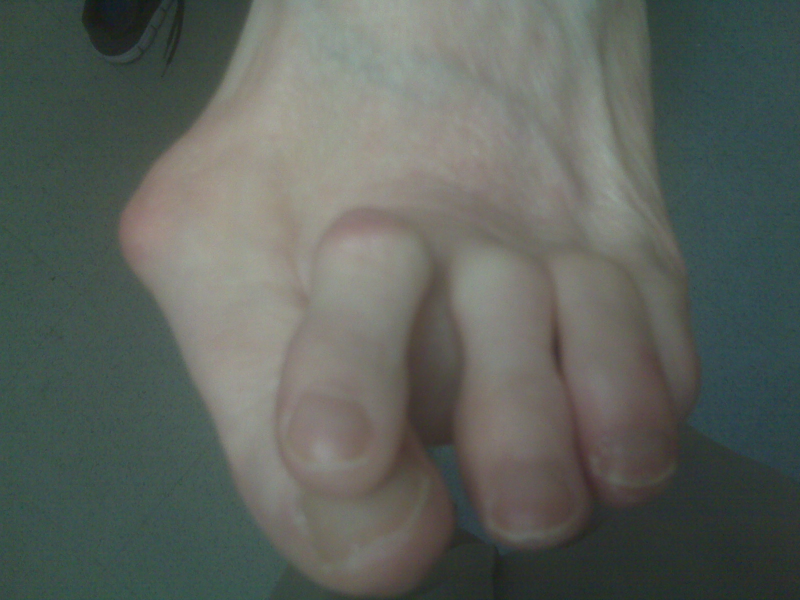
There are about 160 bursae in the human body. These little, fluid-filled sacs cushion pressure and lubricate points between our bones, tendons, and muscles near our joints. The bursae are lined with synovial cells. Synovial cells produce a lubricant that reduces friction. This cushioning and lubrication allows our joints to move easily. When a person has bursitis, inflammation of the bursa, movement or pressure is painful. Overuse, injury and sometimes an infection from gout or rheumatoid arthritis may cause bursitis. Causes Bursitis can be caused by an injury, an infection, or a pre-existing condition in which crystals can form in the bursa. Injury. An injury can irritate the tissue inside the bursa and cause inflammation. Doctors say that bursitis caused by an injury usually takes time to develop. The joints, tendons, or muscles that are near the bursae may have been overused. Most commonly, injury is caused by repetitive movements. Symptoms The following are the most common symptoms of bursitis. However, each individual may experience symptoms differently. Bursitis can cause pain, localized tenderness, and limited motion. Swelling and redness may occur if the inflamed bursa is close to the surface (superficial). Chronic bursitis may involve repeated attacks of pain, swelling, and tenderness, which may lead to the deterioration of muscles and a limited range of motion. The symptoms of bursitis may resemble other medical conditions or problems. Always consult your doctor for a diagnosis. Diagnosis Obtaining a detailed history from the patient is important in diagnosing calcaneal bursitis. The following complaints (which the physician should ask about during the subjective examination) are commonly reported by patients. Other inquiries that the physician should make include the following. The clinician should ask about the patient's customary footwear (whether, for example, it includes high-heeled shoes or tight-fitting athletic shoes). The patient should be asked specifically about any recent change in footwear, such as whether he/she is wearing new athletic shoes or whether the patient has made a transition from flat shoes to high heels or vice versa. Individuals who have been accustomed to wearing high-heeled shoes on a long-term basis may find that switching to flat shoes causes increased stretch and irritation of the Achilles tendon and the associated bursae. The specifics of a patient's activity level should be ascertained, including how far the patient runs and, in particular, whether the individual is running with greater intensity than before or has increased the distance being run. The history of any known or suspected underlying rheumatologic conditions, such as gout, rheumatoid arthritis, or seronegative spondyloarthropathies, should be obtained. Non Surgical Treatment Over-the-counter or custom heel wedges may help to decrease the stress placed on the attachment of the achilles tendon and the associated bursa. If these interventions are ineffective, then some health care providers may inject a small amount of steroids into the bursa. If the condition is associated with Achilles tendonitis, then casting the ankle to prevent motion for several weeks can be effective. Very rarely, surgery may be necessary to remove the inflamed bursa. Surgical Treatment Only if non-surgical attempts at treatment fail, will it make sense to consider surgery. Surgery for retrocalcanel bursitis can include many different procedures. Some of these include removal of the bursa, removing any excess bone at the back of the heel (calcaneal exostectomy), and occasionally detachment and re-attachment of the Achilles tendon. If the foot structure and shape of the heel bone is a primary cause of the bursitis, surgery to re-align the heel bone (calcaneal osteotomy) may be considered. Regardless of which exact surgery is planned, the goal is always to decrease pain and correct the deformity. The idea is to get you back to the activities that you really enjoy. Your foot and ankle surgeon will determine the exact surgical procedure that is most likely to correct the problem in your case. But if you have to have surgery, you can work together to develop a plan that will help assure success.  Overview OverviewIf you sneak a peek at your feet and notice that your toes are crossed, bent, or just pointing at an odd angle, you probably suffer from a Hammer toe. Toes that are scrunched up inside tight shoes or pressed against the toe box of the shoe can bend at the joints and stay that way - resulting in a hammertoe. A hammertoe is a contracture of the toe at one of the two joints in the toe. Due to the pull of the tendons, the joints become more rigid over time. The toe is bent up at the joint and does not straighten out. Causes Risk factors for hammertoe include heredity, a second toe that is longer than the first (Morton foot), high arches or flat feet, injury in which the toe was jammed, rheumatoid arthritis, and, in diabetics, abnormal foot mechanics resulting from muscle and nerve damage. Hammertoe may be precipitated by advancing age, weakness of small muscles in the foot (foot intrinsic muscles), and the wearing of shoes that crowd the toes (too tight, too short, or with heels that are too high). The condition is more common in females than in males.  Symptoms SymptomsHammer toe is often distinguished by a toe stuck in an upside-down ?V? position, and common symptoms include corns on the top of your toe joint. Pain at the top of a bent toe when you put on your shoes. Pain when moving a toe joint. Pain on the ball of your foot under the bent toe. Corns developing on the top of the toe joint. It is advisable to seek medical advice if your feet hurt on a regular basis. It is imperative to act fast and seek the care of a podiatrist or foot surgeon. By acting quickly, you can prevent your problem from getting worse. Diagnosis The treatment options vary with the type and severity of each hammer toe, although identifying the deformity early in its development is important to avoid surgery. Your podiatric physician will examine and X-ray the affected area and recommend a treatment plan specific to your condition. Non Surgical Treatment Conservative treatment is the first choice, often starting with a change of shoes to ones that hammertoe have soft, larger toe spaces. Toe exercises may be prescribed to stretch and strengthen the toe muscles. Over-the-counter straps, cushions or non-medicated corn pads may be recommended to help relieve your symptoms. Surgical Treatment If these treatments are not sufficient at correcting the hammer toe, an operation to straighten the toe may be necessary. This is often performed in conjunction with surgery for a bunion deformity. The surgical treatment of a hammer toe can consist of either cutting the tendons to relieve the pressure that causes the deformity, or fusing the toe so that it points straight permanently.
Overview
 Surgical Treatment If the above simple measures do not make you comfortable, an operation may improve the situation. An operation will not give you an entirely normal foot, but it will correct the deformity of the big toe and narrow your foot back towards a more desirable shape. There are a lot of different operations for bunions, depending on the severity of the deformity, the shape of your foot and whether arthritis has developed in the big toe joint. An orthopaedic surgeon who specialises in foot & ankle surgery can advise you on the best operation for your foot. However, an operation may not make your foot narrow enough to wear tight shoes, nor can it fully restore the strength of the big toe. Overview
During initial contact or heel strike, the ankle rolls inward, causing excessive pronation or flattening of the foot. This action forces the big toe (instead of the ball of foot and all toes) to do all the work to push off the ground. When the ankle overpronates, this causes the tibia (shin bone) to rotate and the femur (thigh bone) to adduct (move toward the mid-line of the body), causing internal rotation or knee valgus (knock-kneed).  Causes In adults, the most common reason for the onset of Over-Pronation is a condition known as Post Tibial Tendonitis. This condition develops from repetitive stress on the main supporting tendon (Posterior Tibial Tendon) of the foot arch. As the body ages, ligaments and muscles can weaken. When this occurs the job of providing the majority of the support required by the foot arch is placed upon this tendon. Unfortunately, this tendon cannot bear the weight of this burden for too long. Eventually it fatigues under the added strain and in doing so the foot arch becomes progressively lower over a period of time. Symptoms With over pronation, sufferers are most likely to experience pain through the arch of the foot. A lack of stability is also a common complaint. Over pronation also causes the foot to turn outward during movement at the ankle, causing sufferers to walk along the inner portion of the foot. This not only can deliver serious pain through the heel and ankle, but it can also be the cause of pain in the knees or lower back as well. This condition also causes the arch to sink which places stress on the bones, ligaments, and tendons throughout the foot. This may yield other common conditions of foot pain such as plantar fasciitis and heel spurs. Diagnosis At some point you may find the pain to much or become frustrated. So what are you options? Chances are your overpronation has led to some type of injury if there's pain. Your best bet is to consult with someone who knows feet. Start with your pediatrist, chiropodist or chiropractor. They'll be able to diagnose and treat the injury and give you more specific direction to better support your feet. One common intervention is a custom foot orthotic. Giving greater structural support than a typical shoe these shoe inserts can dramatically reduce overpronation.  Non Surgical Treatment If a young child is diagnosed with overpronation braces and custom orthotics can be, conjunction with strengthening and stretching exercises, to realign the bones of the foot. These treatments may have to continue until the child has stopped growing, and orthotics may need to be worn for life in order to prevent the foot reverting to an overpronated state. Wearing shoes that properly support the foot, particularly the arch, is one of the most effective treatments for overpronation. Custom-made orthotic inserts can also be very beneficial. They too support the arch and distribute body weight correctly throughout the foot. Motion-control shoes that prohibit pronation can be worn, so may be useful for those with severe overpronation. One good treatment is to walk barefoot as often as possible. Not relying on shoes to support the arch will encourage proper muscle use. Practicing yoga can help to correct poor posture and teach you how to stand with your weight balanced evenly across the whole foot. Surgical Treatment Subtalar Arthroereisis. The ankle and hindfoot bones/midfoot bones around the joint are fused, locking the bones in place and preventing all joint motion. This may also be done in combination with fusion at other joints. This is a very aggressive option usually reserved for extreme cases where no joint flexibility is present and/or the patient has severe arthritic changes in the joint. |
Archives
July 2017
Categories |
 RSS Feed
